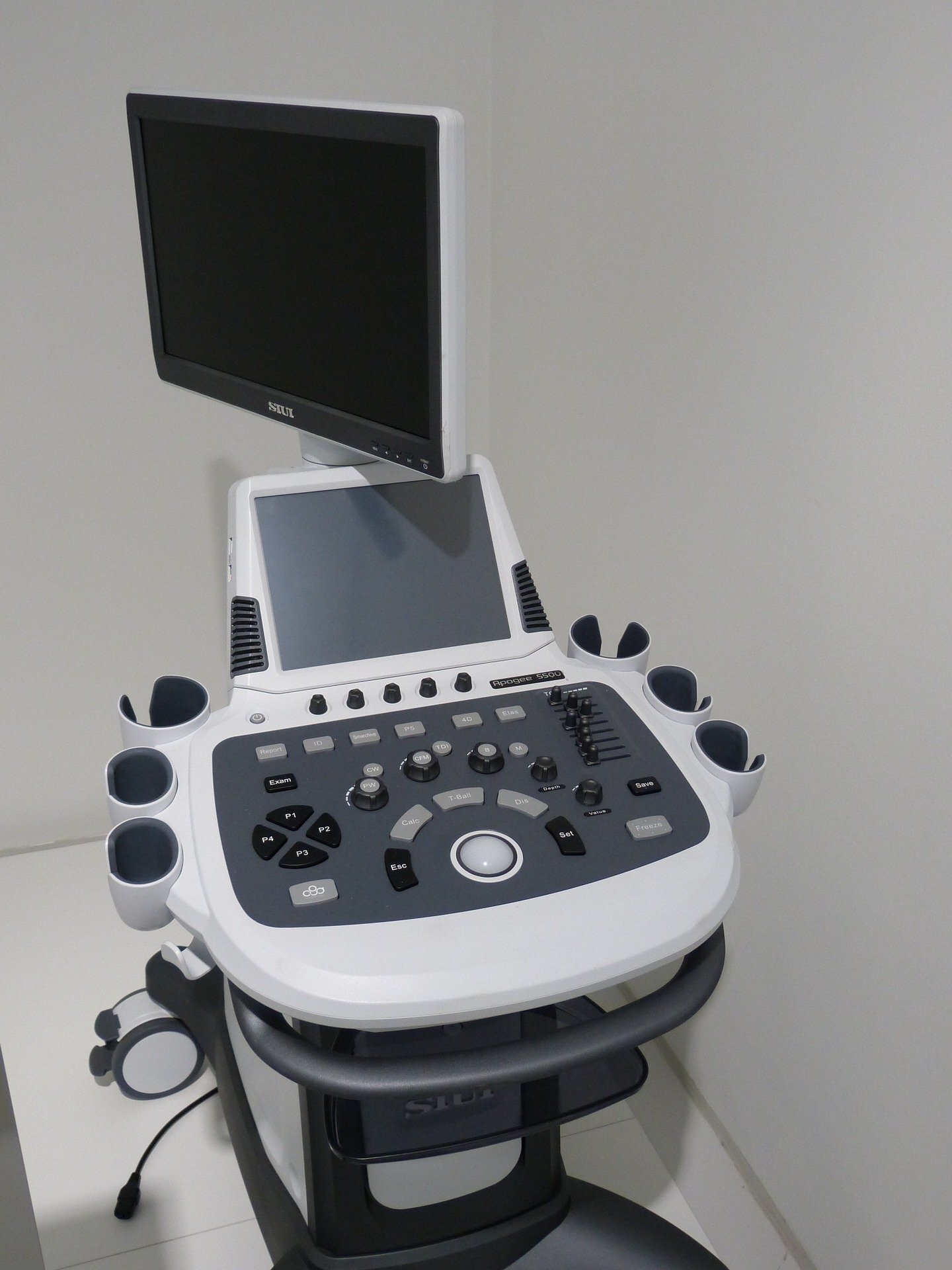
Mayo Clinic researchers have developed an artificial intelligence model that can identify a potentially significant heart obstruction from routine ultrasound videos without relying on specialized Doppler imaging. The study, published in Circulation: Cardiovascular Imaging, focused on patients with hypertrophic cardiomyopathy (HCM), a genetic condition that causes the heart muscle to become abnormally thick.
About two-thirds of HCM patients develop left ventricular outflow tract (LVOT) obstruction, which restricts blood leaving the heart and causes symptoms such as chest pain and shortness of breath. Measuring this obstruction typically requires Doppler echocardiography, which depends on precise ultrasound-beam alignment and operator expertise.
The AI model was trained and tested on 1,833 patients in the Mayo Clinic cohort, with 275 patients used for testing and an additional 46 patients from a hospital in South Korea for external validation. The model used only resting, non-Doppler B-mode ultrasound videos to predict whether a patient had a potentially significant obstruction.
Researchers found that combining information from three standard ultrasound views improved the model's ability to distinguish patients with elevated LVOT gradients. The model also helped identify obstruction that may appear only when the heart is under stress.
The model maintained strong performance in the South Korean validation group despite substantial differences between that population and the patients used to develop the model. In a subset of cases, the AI model identified obstruction more accurately than two expert echocardiographers who reviewed the same non-Doppler images.
The technology is intended to complement, not replace, Doppler echocardiography. By enabling earlier identification of patients with potential LVOT obstruction, it could support timely confirmatory Doppler measurements, stress testing, or referral to an HCM specialty center.
The approach could also support evaluation using portable ultrasound or in settings where comprehensive Doppler assessment may not be readily available, helping expand access to earlier screening and risk assessment. Next steps include additional prospective validation across broader clinical settings, ultrasound platforms, and patient populations.
AI model helps clinicians detect heart obstruction using routine ultrasound images
This is an independent summary. The complete reporting, supporting context and any primary documents remain with Medical Xpress.